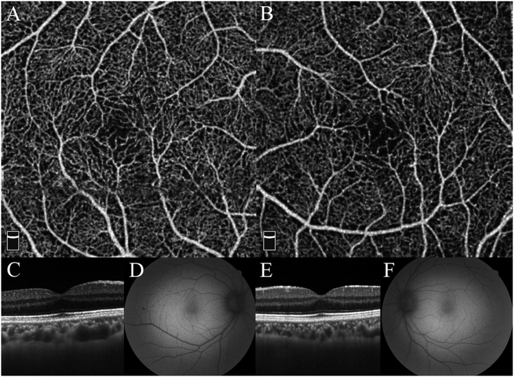
Fig. 1.
Case 1 was a 37-year-old man with normal vision and normal foveal pit. a Optical coherence tomography angiography (OCTA) shows an unmeasurable small foveal avascular zone (FAZ) in the right eye. b Cross-sectional optical coherence tomography (OCT) shows an inner retinal layer, which is seen as a hyperreflective band in spite of the existence of the foveal depression in the right eye. c Fundus autofluorescence shows normal hypo-autofluorescence at the fovea in the right eye. d OCTA image shows the absence of a FAZ in the left eye. e Cross-sectional OCT shows an inner retinal layer, which is seen as a hyperreflective band in spite of the existence of the foveal depression in the left eye. f Fundus autofluorescence shows normal hypo-autofluorescence at the fovea in the left eye

