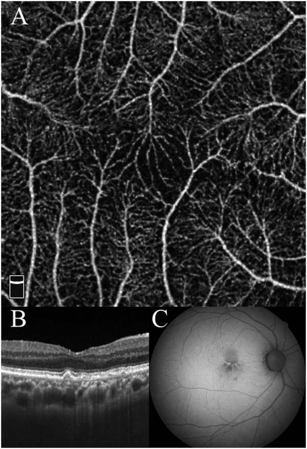
Fig. 3.

Case 3 was a 79-year-old man. a OCTA shows an unmeasurable small size of FAZ. b Cross-sectional OCT image shows a protrusion of the retinal pigment epithelium indicating the presence of soft drusen. A hyperreflective band due to residual inner retinal layers is present. c Fundus autofluorescence shows the linear hyper-autofluorescence due to drusen and normal hypo-autofluorescence at the fovea
