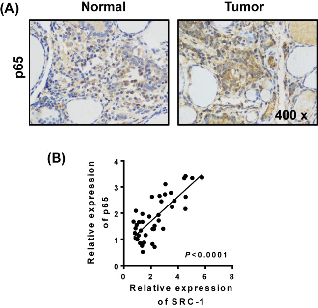
Figure 4. Increased NF-kB signal in thyriod cancer tissues.

The levels of p65 expression in thyroid cancer and relatively normal tissues (n=20) were determined by (A) immunohistochemistry. (B) The correlation between p65 and SRC-1 was analyzed by Q-PCR. P < 0.001; data represent the mean ± S.D.
