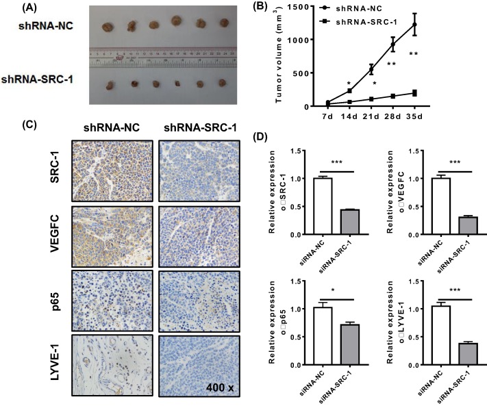
Figure 5. The tumorigenic role of SRC-1 was assessed in vivo.
TPC-1/shRNA-SRC-1 or shRNA-NC cells (2 × 10⁶ per cell type) were subcutaneously injected into the rear flanks of nude mice (six mice per group). (A and B) The mean tumor size in each group (mm3) was analyzed. (C and D) The levels of SRC-1, VEGFC, p65, and LYVE-1 expression were estimated by immunohistochemistry and Q-PCR. *P<0.05, **P<0.001, ***P<0.001; data represent the mean ± S.D.

