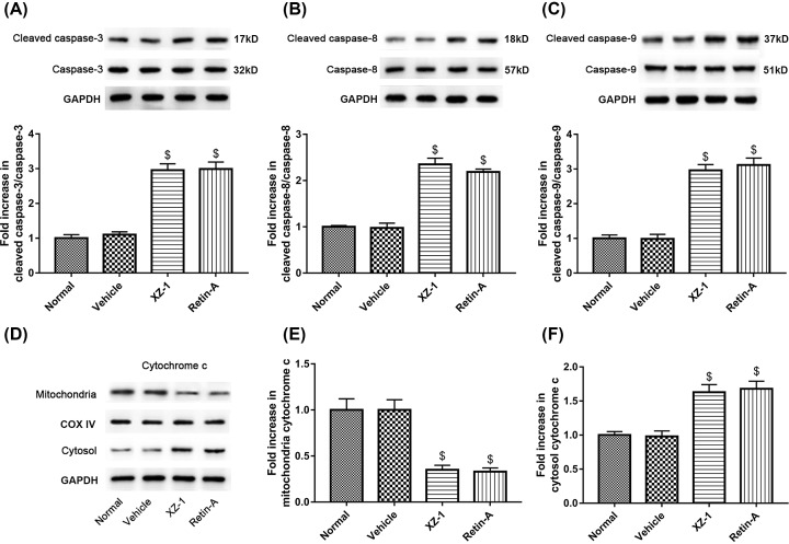
Figure 3. Cleaved caspases (-3, -8, and -9) and cytochrome c expression in the pyloric area of the stomachs from GED rats.
The protein expression of (A) cleaved caspase-3 and caspase-3, (B) cleaved caspase-8 and caspase-8, and (C) cleaved caspase-9 and caspase-9 in the pyloric area of the stomachs from rats in each group was evaluated by Western blot. GAPDH served as the loading control. The quantitative analysis of their blots was shown below. (D) The separated cytoplasmic and mitochondrial protein isolated from total proteins in the pyloric area of the stomachs from rats in each group was subjected to Western blot analysis of cytochrome c. COXIV served as the subcellular marker for mitochondrial cytochrome c and GAPDH for cytosolic cytochrome c. (E) The quantitative analysis of protein expression of mitochondrial cytochrome c was shown. (F) The quantitative analysis of protein expression of cytosolic cytochrome c was shown. $P<0.05 compared with Vehicle group. n=15 per group.

