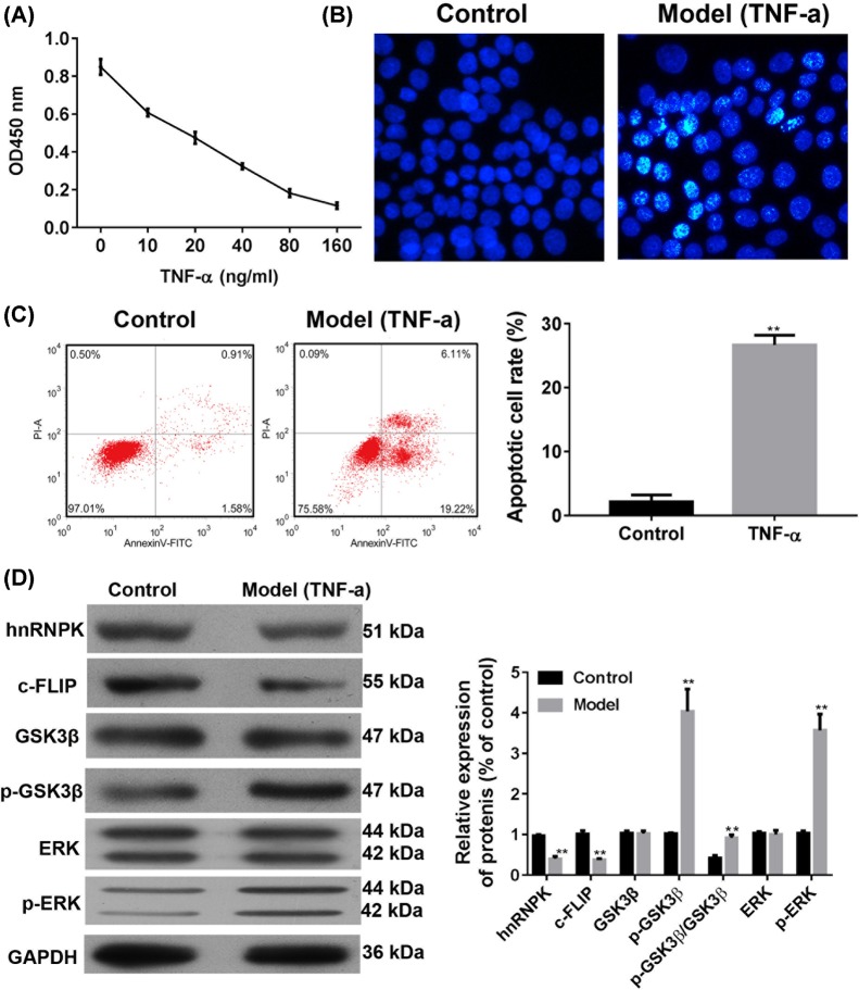
Figure 2. Effects of TNF-α on cell apoptosis and protein expressions.
(A) Primary podocytes were seeded in 96-well plates and were treated with TNF-α for 24 h, then the viability was measured by CCK8 assay. (B) After treatment with 5 ng/ml TNF-α for 24 h, apoptotic morphological changes in the primary cells were examined by Hoechest staining under fluorescence microscopy. (C) The apoptosis of primary podocytes were measured by flow cytometry. (D) Expressions of hnRNP K, c-FLIP, GSK3β, p-GSK3β, ERK, and p-ERK were analyzed by Western blot analysis. **P<0.01, compared with the control group by two-tailed Student’s t test. Magnification = 400×.

