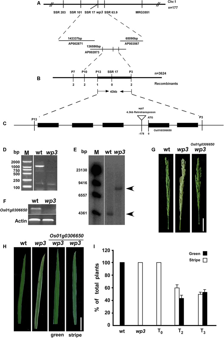
FIGURE 3.
Map-based cloning of WP3. (A) Genetic mapping of WP3 using SSR markers on the long arm of chromosome 1. BAC clones covering the corresponding region were shown. (B) Fine mapping of WP3. Number of recombinants was indicated below each marker. (C) Distributions of five predicted genes and schematic representation of WP3 structure. The predicted translation start site (ATG) of WP3 and site of retrotransposon insertion (inverted triangle) were indicated. (D) PCR amplification of WP3 promoter region. M indicated the DNA ladder used. (E) Southern blotting in wild type and wp3 plant. Genomic DNA from wild type and wp3 plant was prepared, digested by EcoR I, transferred to membranes, and probed by a 32p-labeled DNA fragment containing WP3 ORF. (F) RT-PCR amplification of WP3 in wild type rice and wp3 mutant. Actin was amplified as an internal control. (G,H) Representative images of panicle (G) and leaf (H) from wild-type, wp3 and a transgenic wp3 plant with expression of WP3. Scale bars = 5 cm. (I) Quantification of plants with white-stripe leaves in wild type, wp3, and WP3 expressed wp3 mutant at various generations.

