FIGURE 5.
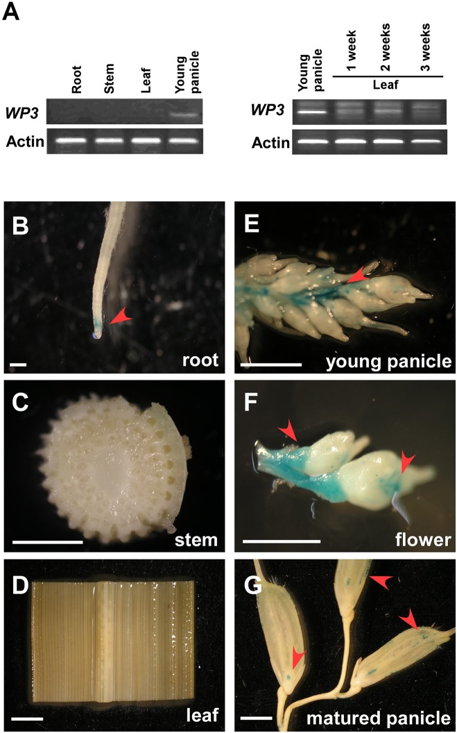
Expression patterns of WP3. (A) RT-PCR amplification of WP3 in various rice tissues. Actin was used as a internal control. The experiments were performed in three biological replicates and representative results were shown. (B–G) WP3 promoter driven GUS expression in rice root (B), stem (C), leaf (D), young panicle (E), flower (F), and matured panicles (G). Arrows indicated the expression of GUS. Scale bars = 0.2 cm.

