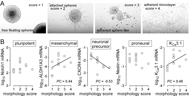
Fig. (1).
Heterogeneity of primary glioblastoma cultures. A, B. Morphological phenotypes of primary glioblastoma cultures (A) and their association (B) with mRNA abundances of stem cell markers and of IKCa3.1 (PC: Pearson correlation coefficient). Resected glioblastoma tissue (patients gave informed consent and this study has been approved by the local ethic committee under project #579/2015BO2) was dissociated enzymatically and mechanically and plated in Complete NeuroCult™ NS-A Proliferation medium containing 20 ng/ml rhEGF, 10 ng/ml rhbFGF and 0.0002% heparin (STEMCELL Technologies Germany GmbH, Cologne, Germany). Every second day 1/10 of the original medium volume was replenished (for RT-PCR, see Legend to Fig. 2).

