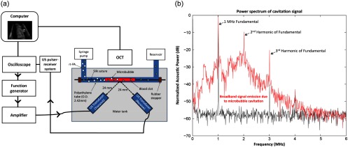
Fig. 1.
In vitro experimental setup and broadband signal emission. (a) A polyethylene tube containing a fixed blood clot was prepared and placed on a custom microscope stage with an acoustic chamber fitted with two ultrasound transducers. Optical coherence tomography was employed to image the thrombi over 30 min of ultrasound treatment. (b) The broadband signal emission, which indicates the occurrence of MB destruction, was evident when MBs were present in the perfusate (red, top) but not when ultrasound was applied without MBs (black, bottom).

