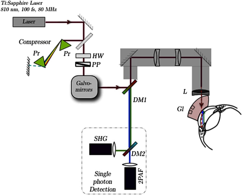
Fig. 1.
Schematic of the custom multiphoton gonioscopy microscope. The pulsed femtosecond laser beam is first sent through a prism compressor and passed through two galvanometric scanning mirrors for raster scanning at the sample. A custom-made lens relay system is used to convert our inverted microscope (Olympus IX71) to an upright microscope for in situ imaging. The multiphoton microscopy (MPM) signal from the sample is collected back through the microscope objective and separated from the excitation laser light with a dichroic mirror (DM1). An additional dichroic mirror (DM2) is used to spectrally separate the second-harmonic-generation (SHG) and two-photon autofluorescence (2PAF) signals, which are subsequently focused on two single-photon counting photodetectors for data acquisition. Pr, prism; HW, half-wave plate; PP, polarizer; Gl, gonioscopic lens; L, focusing lens.

