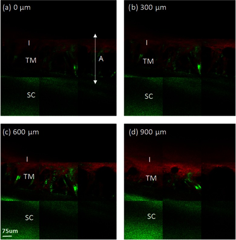
Fig. 2.
Acquired MPM images of the TM of a porcine eye taken through a gonioscopic lens. Panels 2(a)–2(d) are tiles of combined MPM images of the porcine trabecular meshwork (TM) at different depths, where 2(a) is closest to the anterior chamber and 2(d) is farthest away from the anterior chamber. The images are 300 µm apart. SHG signal is displayed in green and 2PAF is displayed in red. A collagen-rich mesh structure (SHG) is observed in all images. The mesh structure is located in a gap that separates two distinct regions: a strong SHG region (sclera) and a strong 2PAF region (iris). SC, sclera; I, iris; A, irido-corneal angle.

