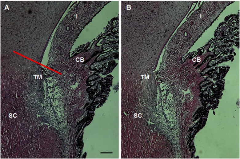
Fig. 4.
MPM gonioscopy imaging of the TM region of the porcine eye does not appear to damage ocular structures. A histological section of the region of the porcine eye imaged by MPM (b) shows no distortion or photocoagulation of the tissues near the drainage angle compared to a region from the opposite side of the eye (a). The red line in 4(a) shows the approximate imaging plane of Fig. 2(a); 2(b) and 2(c) would be located parallel to this plane traveling deeper into the scleral/TM region. CB, ciliary body. The scale bar represents 200 µm.

