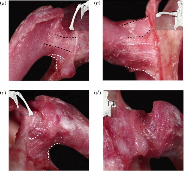
Figure 2.
Images of the hip joint capsule and its ligamentous thickenings in the right hip of a common quail: (a) anterior, (b) ventral, (c) posterior and (d) dorsal views. Thumbnails in each view display the approximate hip pose at which each image was taken. Black dashed lines indicate the borders of the pubofemoral ligament; white dashed lines indicate the approximate borders of the ischiofemoral ligament. (Online version in colour.)

