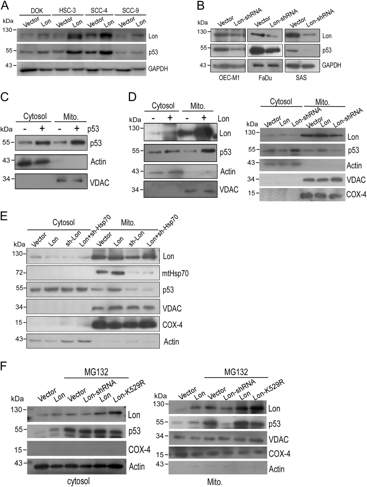
Fig. 5. Chaperone Lon–mtHsp70 is required for mitochondrial p53 accumulation that is important to prevent the cytosolic distribution of p53 from proteasome-dependent degradation in cancer cells.
a, b Mitochondrial Lon is important for the stability/level of p53 protein in cancer cells. For overexpression experiment, oral cancer cells were transfected with the plasmid encoding Myc-tagged Lon. For knocking down experiment, Lon expression was inhibited by Lon-shRNA transfection. Immunoblotting were performed using the indicated antibodies. c Immunoblotting analysis of increased p53 in mitochondrial fraction. p53 was overexpressed in OEC-M1 cells by transfected with the plasmid encoding p53. Immunoblotting were performed using the indicated antibodies. The purity of each cell fractions was monitored by immunoblotting for cytoplasmic (Actin) and mitochondrial (VDAC) markers. d Chaperone Lon is required for mitochondrial p53 accumulation. OEC-M1 cells were transfected with the plasmid encoding Myc-tagged Lon or Lon-shRNA. Immunoblotting were performed using the indicated antibodies. The purity of each cell fractions was monitored by immunoblotting for cytoplasmic (Actin) and mitochondrial (VDAC and COX-4) markers. e Chaperone Lon-induced mitochondrial p53 accumulation is dependent on mtHsp70. OEC-M1 cells were transfected with the plasmid encoding Myc-tagged Lon, Lon-shRNA, and/or mtHsp70-shRNA. Immunoblotting were performed using the indicated antibodies. The purity of each cell fractions was monitored by immunoblotting for cytoplasmic (Actin) and mitochondrial (VDAC and COX-4) markers. f The chaperone activity of Lon is required for mitochondrial p53 accumulation and the prevention of the cytosolic distribution of p53 from proteasome-dependent degradation. OEC-M1 cells were transfected with the plasmid encoding WT-Lon, Lon-shRNA, or the ATPase mutant of Lon (LonK529R). The transfected cells were treated with 10 μM MG132 for 2 h. Immunoblotting were performed using the indicated antibodies. The purity of each cell fractions was monitored by immunoblotting for cytoplasmic (Actin, left) and mitochondrial (VDAC and COX-4, right) markers

