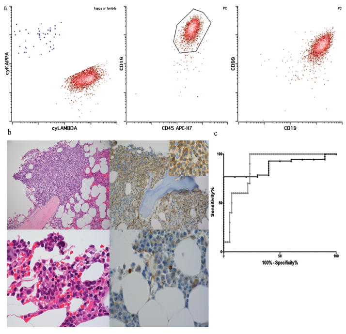
Figure 5.
A. Plasma cells in B cell lymphoma showed bright CD99 expression. Representative example is shown. Lambda-restricted abnormal plasma cells (red) from a patient with LPL showed bright CD99 expression.
B. CD99 expression could also be detected plasma cells in lymphoma with plasma cell component by immunohistochemistry (top row, H&E 400X left CD99 400X and 1000X(inset), right). In contrast CD99 was usually absent in myeloma by immunohistochemistry (bottom row, H&E 1000X left, CD99 1000X, right).
C. Receiver-operator characteristic curves for myeloma vs plasma cells from lymphoma are shown for all neoplastic samples (triangles, grey) and those without CCND1 translocation (circles, black).

