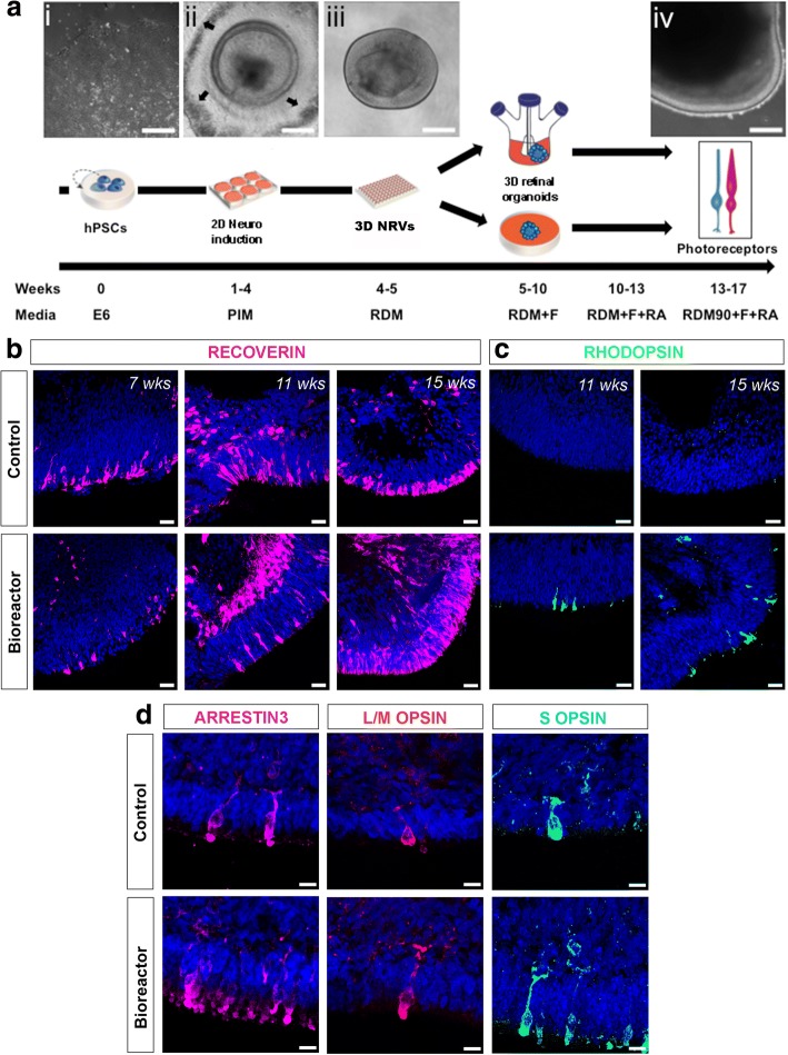
Fig. 1.
Photoreceptor maturation following bioreactor differentiation. a Schematic showing different stages of differentiation protocol. Representative phase-contrast images: (i) hPSCs at day 0 of differentiation; (ii) developing neuroepithelia surrounded by RPE cells (black arrows) at week 4; (iii) isolated retinal cup at week 5; (iv) mature retinal organoid at week 17. Following retinal cup formation, samples cultured in 100-mm cell culture plates or 100-ml bioreactors to form retinal organoids. Mature photoreceptors observed between days 90 and 120. b, c RECOVERIN and RHODOPSIN-positive photoreceptor cells in both control and bioreactor conditions at indicated number of weeks of differentiation. d Immunohistochemistry of L/M-OPSIN, Cone Arrestin (ARRESTIN3) and S-OPSIN cone markers at week 16 of differentiation in control and bioreactor conditions. Scale bars: 20 μm (a), 25 μM (b, c), 10 μM (d). hPSC human pluripotent stem cell, NRV neuroretinal vesicle, RDM + F RDM supplemented with 10% foetal bovine serum + 2% Glutamax + 100 μM taurine, RA retinoic acid

