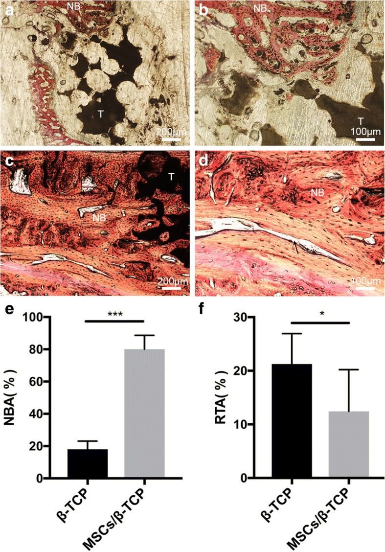
Fig. 8.

Histology and histomorphology evaluation. Van Gieson staining of hard tissue slices 6 months after operation from pure porous β-tricalcium phosphate (TCP) treatment (a,b) and mesenchymal stem cells and porous β-tricalcium phosphate composites (MSCs/β-TCP) (c,d) treatment of 3-cm bone defects. Black arrow indicates new Haver’s tube formation. e,f Percentage of new bone area (NBA) and residual β-TCP area (RTA). NB new bone tissues, T residual β-TCP. *p < 0.05, ***p < 0.01
