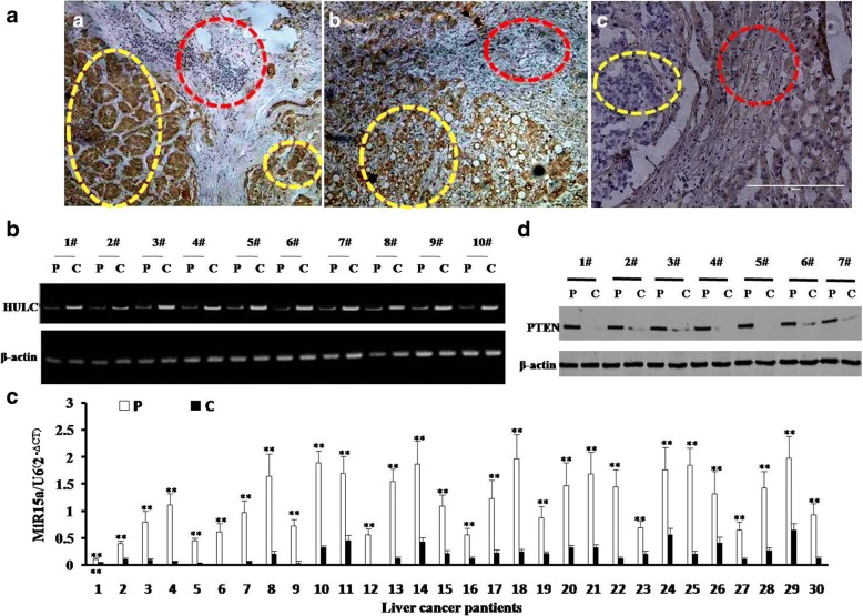
Fig. 1.
Expression analysis of HULC, miR15a, PTEN in human liver cancer tissue. a (left&middle) The representative analytic results of in situ hybridization for HULC and (right) immunohistochemistry staining for PTEN in human liver cancer tissue and their paired adjacent noncancerous tissues from the same patient (DAB stainning, original magnification× 100). b The representative analytic results of RT-PCR with HULC primers in liver cancer tissue (c) and its paracancerous liver tissues (p) respectively. β-actin as internal control. c The Real-time RT-PCR results of mature miR15a in liver cancer tissue (c) and its paracancerous liver tissues (p), respectively. U6 as internal control. d The representative analytic results of western blotting with anti-PTEN in liver cancer tissue (c) and its paracancerous liver tissues (p), respectively. β-actin as internal control

