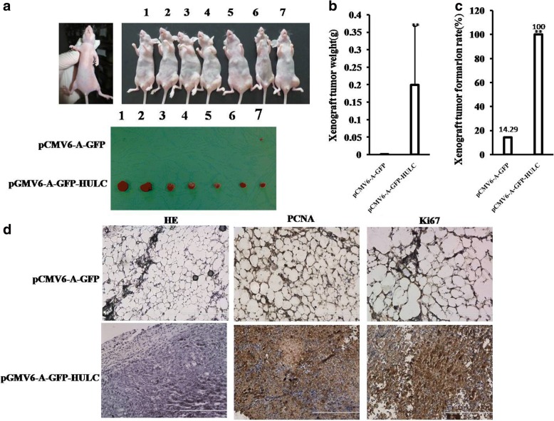
Fig. 3.
HULC promotes liver cancer cell growth in vivo. a The photography of xenograft tumors from Balb/C null mouse injected with Hep3B cells transfected with pCMV6-A-GFP,pCMV6-A-GFP-HULC subcutaneously at armpit. b The xenograft tumors weight (gram) in the two groups indicated in left. Data were means of value from nine Balb/c mice, mean ± SEM, n = 7,*, P < 0.05;**, P < 0.01. c The xenograft tumors formation rate(%) in the two groups indicated in left. d Hematoxylin-eosin (HE) staining of xenograft tumors (original magnification× 100). And anti-PCNA and anti-k67 immunostainningin xenograft tumor samples. (original magnification× 100)

