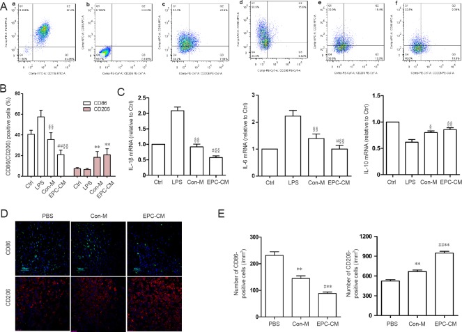
Figure 1.
Effects of EPC-CM on inflammatory cytokine levels in vitro and in vivo.
(A) Representative flow cytometry data showing the effect of EPC-CM on BMDMs. (a) Mature BMDMs were defined as CD11b+/F4/80+ subpopulations (upper right), with the purity displayed as percentage of the parent population. (b) Control BMDMs were incubated with CD11b and F4/80 antibody and used to set up the gate. (c) Control BMDMs without LPS stimulation were incubated with anti-rat CD11b, F4/80, CD86 and CD206 antibodies. M1 macrophages are CD11b+/F4/80+/CD86+/CD206− (Q1), whereas M2 macrophages are CD11b+/F4/80+/CD86−/CD206+ (Q3). (d) BMDMs treated with LPS. (e) BMDMs cultured with Con-M and simultaneously stimulated with LPS. (f) BMDMs cultured with EPC-CM and stimulated with LPS. (B) Quantitation of M1 and M2 cells among the different groups. Compared with the Con-M group, EPC-CM significantly reduced M1 activation, while M2 cells remained relatively unchanged. (C) mRNA expression levels of inflammatory cytokines (optical density ratio) among groups. (D) Immunofluorescence staining for CD86 (M1 marker) and CD206 (M2 marker) in the epicenter 7 days after SCI (n = 5 per group; green: CD86; red: CD206; blue: DAPI). (E) Quantification of CD86- and CD206-positive cells at 7 days after SCI. **P < 0.01, vs. PBS group; #P < 0.05, ##P < 0.01, vs. Con-M group. §P < 0.05, §§P < 0.01, vs. LPS. Data are presented as the mean ± SD (one-way analysis of variance followed by the least significant difference post hoc test). The experiment was performed at least three times. Ctrl: Control; PBS: phosphate-buffered saline; EPC-CM: endothelial progenitor cell–conditioned medium; Con-M: control medium; BMDMs: bone marrow-derived macrophages; LPS: lipopolysaccharide; IL: interleukin; DAPI: 4′,6-diamidino-2-phenylindole.

