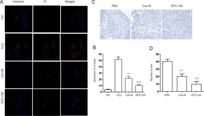
Figure 2.
Effect of EPC-CM on apoptosis in vitro and in vivo.
(A) In vitro representative fluorescence images of Hoechst and PI double staining (original magnification, 200×; blue: Hoechst 33258; red: PI). (B) Quantitative assessment of apoptotic cells in the various groups. EPC-CM significantly reduced H2O2-induced spinal cord neuron apoptosis. The experiment was performed at least three times. (C) Cells in the gray matter in the epicenter stained with TUNEL one week after injury (in vivo). The nuclei of TUNEL-positive (apoptotic) cells are stained a dark color (n = 5 per group; original magnification, 200×). (D) Quantitative analysis of TUNEL-positive cells. Compared with the PBS group, the EPC-CM and Con-M groups had significantly fewer TUNEL-positive cells. **P < 0.01, vs. PBS group; #P < 0.05, ##P < 0.01, vs. Con-M group. ††P < 0.01, vs. H2O2 group. Data are presented as the mean ± SD (one-way analysis of variance followed by the least significant difference post hoc test). The experiment was performed at least three times. Ctrl: Control; PBS: phosphate-buffered saline; PI: propidium iodide; EPC-CM: endothelial progenitor cell-conditioned msedium; Con-M: control medium; TUNEL: terminal deoxynucleotidyl transferase dUTP nick-end labeling.

