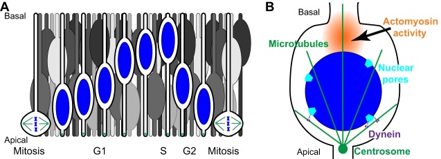
Fig. 3.
Interkinetic nuclear migration. (A) Illustrated here are nuclei (blue) that undergo interkinetic nuclear migration within a pseudostratified epithelium. Cells with blue nuclei are organized chronologically to show one cell cycle. Other cells (in gray) illustrate the crowded nature of this epithelium. As G2-phase nuclei actively move towards the apical side, they passively push G1-phase nuclei out of the way, resulting in their migration towards the basal surface. (B) Active apical movement during G2 phase; dynein (purple) is recruited to the nuclear surface at nuclear pores (light blue) to move nuclei on microtubules (green). Actomyosin contracts in a zone (orange) behind the nucleus to push the nucleus to the apical surface of the epithelium just prior to mitosis.

