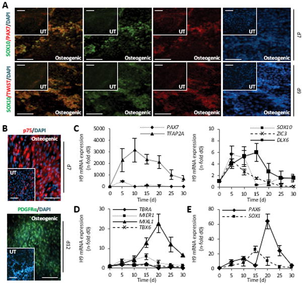
Figure 3. Intermediate differentiation in H9 cells is through the neural crest lineage.

(A, B) Double staining with antibodies against SOX10/PAX7, SOX10/TWIST1, and single stains with p75NTR, and PDGFRα at indicated time points of differentiation. Scale bar = 100 μm.
(C–E) Quantitative mRNA analysis of early lineage genes, normalized to GAPDH, n=3 independent samples ± SD.
T/BRA, T-Brachyury; GAPDH, glyceraldehyde 3-phosphate dehydrogenase.
