
Figure 1.
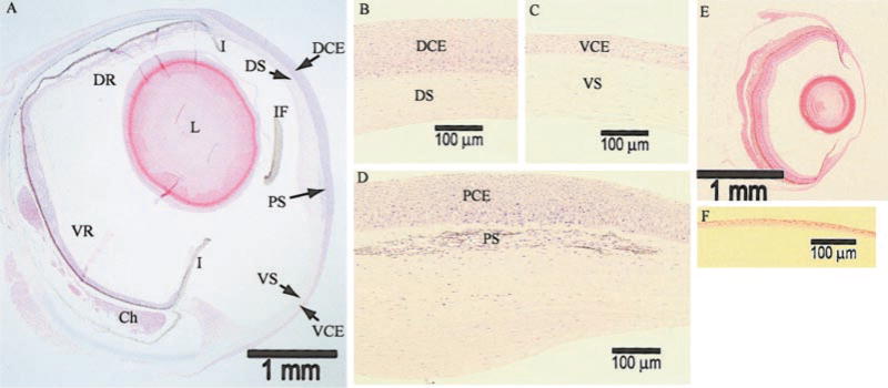
Light microscopy of Anableps and zebrafish eyes. A) Bright-field microscopy of 2 μm-thick methacrylate-embedded sections stained with hematoxylin and eosin at 25× magnification. DCE, dorsal corneal epithelium; VCE, ventral corneal epithelium; PCE, pigmented corneal epithelium; PS, pigmented strip; I, iris; IF, iris flap; DS, dorsal corneal stroma; VS, ventral corneal stroma; DR, dorsal retina; VR, ventral retina; Ch, choroidal tissue; L, lens. B) Dorsal cornea, at 200× magnification. C) Ventral cornea, at 200× magnification. D) Central cornea bisected by the water surface, at 200× magnification. E) Zebrafish eye, at 25× magnification. F) Zebrafish cornea, at 200× magnification. Scale bars are provided for reference.