
Fig. 2.
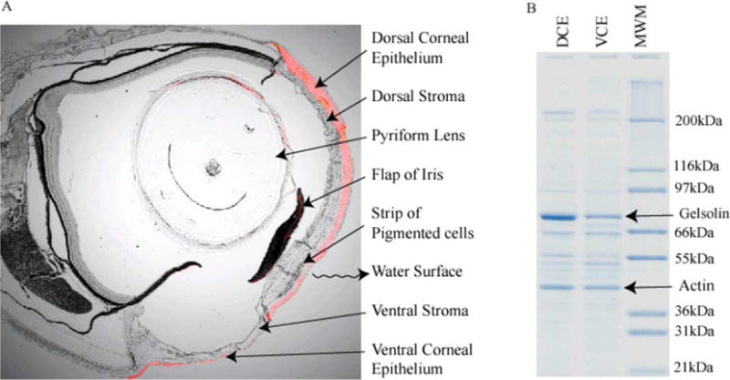
Adaptations in the structure and water-soluble protein compositions of Anableps corneas. (A) Superimposition of bright field photomicrograph with fluorescence image of immunohistochemistry with anti-gelsolin antibodies (red color). Magnification: 25 ×. (B) Profile of water-soluble proteins from the dorsal corneal epithelial cells (DCE) and the ventral corneal epithelial cells (VCE) of Anableps. Gelsolin and actin are indicated.