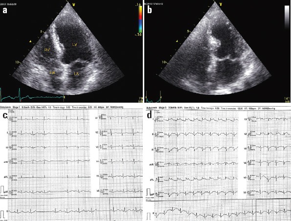
Figure 1.

Baseline TTE showed no wall motion abnormality in a 4-chamber view with LVEF of 55% (panel A), and a concomitant ECG showed a sinus rhythm with SA arrest and no ST-T change (panel C). TTE at peak of the test in 4-chamber view showed akinetic myocardium from the mid to apical part of septum with LVEF of 25% (panel B), and a concomitant ECG showed an ST elevation in I, aVL, V2 with subtle ST depression in inferior leads at peak of test (panel D). ECG -electrocardiogram; LA - left atrium; LV - left ventricle; LVEF - left ventricle ejection fraction; RA - right atrium; RV - right ventricle; SA - sinoatrial node; TTE - transthoracic echocardiogram
