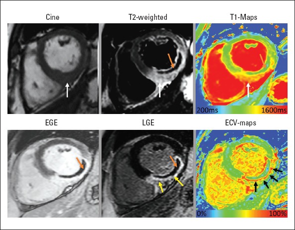
Figure 2.

In vivo histopathology assessment by multi-parametric cardiac magnetic resonance imaging (MRI) of a patient who presented with acute ST-elevation myocardial infarction. On cine images, inferio-lateral segments demonstrate increased thickness secondary to myocardial edema. T2-weighted imaging (white arrow) and T1 maps (white arrow) confirm the presence of myocardial edema. A hypointense core is seen on T2-weighted imaging, which is consistent with the diagnosis of intra-myocardial hemorrhage (orange arrow). This is also present in EGE imaging and LGE imaging confirming the presence of MVO. LGE imaging confirms large infero-lateral MI and MVO. ECV maps demonstrate >60% ECV in the scar tissue, suggesting severe tissue damage (black arrows). Also, note that IMH/MVO within the infarct results in pseudo-normalization of native T1 and ECV
