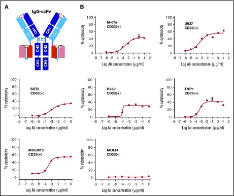
Figure 1.
Anti-CD33 BsAb BC133 lysed AML cells in vitro at femtomolar EC50. (A) A schematic diagram of anti CD33×CD3 huM195 BsAb named BC133 in IgG(L)-scFv format. Heavy chains and light chains are shown in dark and light colors, respectively. (B) T-cell–mediated cytotoxicity against various CD33+ human AML cell lines in the presence of BC133 was assessed by a 4-hour chromium release assay. CD33– MOLT4 cells were used as negative control.

