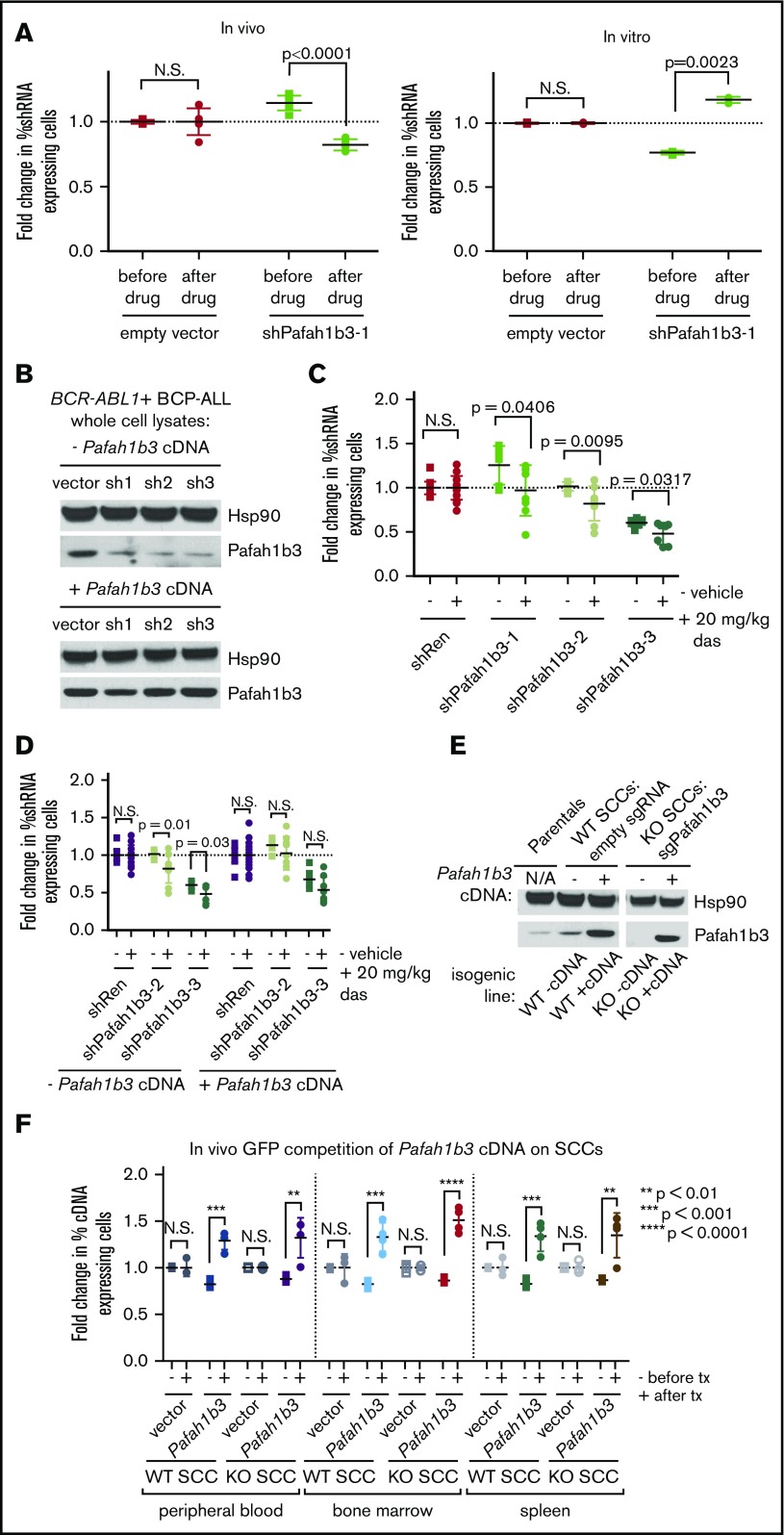
Figure 3.
Pafah1b3 loss confers sensitivity to dasatinib specifically in vivo. (A) Scatterplots showing that the initial hairpin against Pafah1b3 from the 20K screen depletes significantly after dasatinib (das) treatment (20 mg/kg q.d. for 3 days starting 11 days postinjection of 106 BCR-ABL1+ BCP-ALL cells; 1 nM in culture for 3 days) in vivo (left) but not in vitro (right) in GFP competition assays performed as described in supplemental Figure 6; fold changes are normalized to empty vector. Values are an average of at least 7 mice per condition. (B) Western blot showing Pafah1b3 knockdown by shRNAs in the presence of absence of a Pafah1b3 cDNA in cultured BCR-ABL1+ BCP-ALL cells infected with cDNA and/or shRNA and sorted by flow cytometry or selected by antibiotic resistance for pure populations. shPafah1b3-1 is the initial shRNA identified in 20K screen that targets an exon; shPafah1b3-2 and shPafah1b3-3 are additional shRNAs targeting the 3′UTR of the Pafah1b3 gene. Hsp90 is used as a loading control; Hsp90 and Pafah1b3 panels of the same lysates are different exposures of the same blot. −/+ cDNA lysates were run on distinct blots. (C) Scatterplot showing fold change in percentage of shPafah1b3-expressing cells from transplant to morbidity in both untreated and dasatinib-treated (20 mg/kg, 3 days q.d. starting 11 days postinjection of 106 BCR-ABL1+ BCP-ALL cells) mice. GFP competition is performed as described in supplemental Figure 6, except that some mice are treated with vehicle instead of dasatinib, and no pretreatment sample is taken. − indicates vehicle-treated mice (morbidity/transplant); + indicates dasatinib-treated mice (morbidity/transplant). Fold changes are normalized to shRenilla; values are an average of at least 7 mice per condition. (D) Scatterplot showing fold change in percentage of shPafah1b3-expressing cells that coexpress either the Pafah1b3 cDNA or an empty GFP vector from transplant to morbidity in both untreated and dasatinib-treated (20 mg/kg, 3 days q.d. starting 11 days postinjection of 106 BCR-ABL1+ BCP-ALL cells) mice. − indicates vehicle-treated mice (morbidity/transplant); + indicates dasatinib-treated mice (morbidity/transplant); the presence or absence of the Pafah1b3 cDNA is noted below the x-axis. Fold changes are normalized to shRenilla vector for matching cDNA condition. GFP competition assays are performed as in panel C. Values are an average of at least 7 mice per condition. (E) Western blot showing Pafah1b3 expression in single-cell clones (SCCs) generated by using CRISPR/Cas9 empty constructs (WT −cDNA, WT +cDNA) or CRISPR/Cas9 constructs targeting the Pafah1b3 gene (KO −cDNA, KO +cDNA), with or without rescue of Pafah1b3 protein levels via expression of the Pafah1b3 cDNA. Each pair of isogenic lines was created from a single SCC. Hsp90 is used as a loading control. Hsp90 and Pafah1b3 panels of the same lysates are different exposures of the same blot; WT vs KO SCCs are from the same exposure(s) of the same blot, but are edited to remove clones that were not used in subsequent experiments. (F) Scatterplot showing fold change in cDNA-expressing cells on either Pafah1b3 WT or KO backgrounds before and after dasatinib treatment (20 mg/kg, 3 days q.d. starting 7 days postinjection of 106 BCR-ABL1+ BCP-ALL cells) in mouse PB, BM, and SPL, as normalized to an empty vector control. Pre- and posttreatment samples are taken from distinct mice rather than longitudinally sampled. − indicates fold change before treatment (pre/transplant); + indicates fold change after treatment (post/pre). Fold changes are normalized to empty vector control within each organ for the same SCC (WT or KO). GFP competition assays are performed as described in supplemental Figure 6, except timing of treatment is changed as noted, and mice are not longitudinally sampled. Values are an average of at least 3 mice per condition and time point. Error bars in all panels indicate standard deviation; P values were calculated using the Student t test.

