Figure 4.
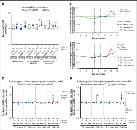
Changes to Pafah1b3 levels alter distribution of leukemia cells in vivo after dasatinib therapy. (A) Scatterplots showing fold change in cDNA-expressing cells on either Pafah1b3 WT or KO backgrounds before and after dasatinib treatment (1 nM for 3 days) in vitro when BCR-ABL+ BCP-ALL cells are cultured alone, with BM–derived stromal cells, or with SPL-derived stromal cells. − indicates fold change before treatment (pre/plating); + indicates fold change after treatment (post/pre). Fold changes are normalized to an empty vector control from the same culture condition and SCC. (B) Plots showing the fold change in Pafah1b3 cDNA-expressing cells, as compared with percentage of cDNA-expressing cells at transplant, over time in the BM and SPL on both Pafah1b3 WT (top) and Pafah1b3 KO (bottom) backgrounds over the course of dasatinib treatment (20 mg/kg, 3 days q.d. starting 7 days postinjection of 106 BCR-ABL1+ BCP-ALL cells; vertical gray lines indicate time period when dasatinib is present). Asterisks denoting significance are color-matched to the organs; changes are not significant if not otherwise noted. (C-D) Scatterplots show relative enrichment of Pafah1b3 cDNA-expressing cells, or cells expressing an empty vector control, on a Pafah1b3 WT or KO background from transplant to the start of treatment (pre/transplant) vs relative enrichment from (C) transplant to morbidity (morbidity/transplant) or (D) transplant to 5 days posttreatment (5 days post/transplant) from GFP competition assays shown in panel B in PB, BM, and SPL. − indicates fold change before treatment; + indicates fold change after treatment. Fold changes are normalized to empty vector control from the same organ and SCC. Relative enrichment is defined as the fold change of percentage of cDNA+ cells in each organ normalized to the fold change of percentage of cDNA+ cells in the blood of the same mouse. For all panels, values are an average of at least 3 mice per genotype at each time point. Individual mice are used for each time point. GFP competition assays are performed as described in “Methods”/supplemental Figure 6, except for timing of treatment as detailed here. Error bars represent standard deviation; P values were calculated using the Student t test.

