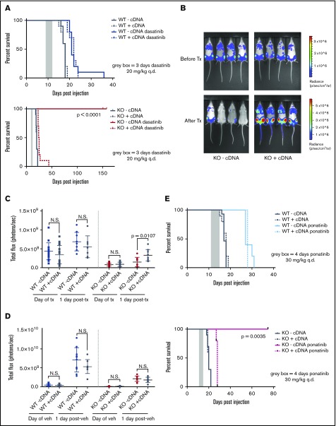
Figure 5.
CRISPR/Cas9-mediated KO of Pafah1b3 in BCR-ABL1+BCP-ALL cells results in increased survival of leukemia-bearing mice after TKI treatment. (A) Survival analysis of dasatinib-treated mice receiving 104 WT-cDNA or WT+cDNA cells (top) or KO −cDNA or KO +cDNA cells (bottom). Significance was calculated using the Mantel-Cox test; the gray rectangle indicates the time period (3 days q.d. starting at 9 days postinjection) over which dasatinib was administered at 20 mg/kg. Ten mice per condition were used in 2 independent experiments. (B) In vivo luminescent imaging of representative mice before and after dasatinib treatment of mice from survival curve shown in panel A that received KO −cDNA or KO +cDNA cells. Different color scales are used in before treatment and after treatment images to for visualization purposes, but images at each time point use the same color scale and duration of exposure. Bioluminescent images were collected using a Xenogen IVIS system and analyzed using Living Image version 4.4 software (Caliper Life Sciences). (C-D) Scatterplots quantifying in vivo luminescent imaging data (total flux = photons per second) before and after dasatinib treatment (C) or vehicle treatment (D) of mice from survival curves shown in panel A. Values are an average of at least 8 mice per genotype. Error bars indicate standard deviation; P values were calculated using the Student t test. (E) Survival analysis of ponatinib-treated mice receiving 104 WT −cDNA or WT +cDNA cells (top) or KO −cDNA or KO +cDNA cells (bottom). Significance was calculated using the Mantel-Cox test; the gray rectangle indicates the time period (4 days q.d, starting at 11 days postinjection) over which ponatinib was administered at 30 mg/kg. Five mice were used per condition.

