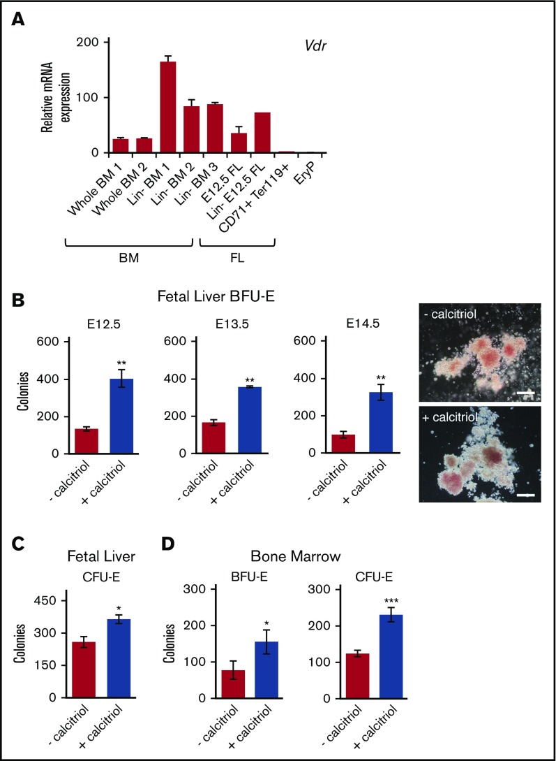
Figure 1.
Activation of Vdr signaling stimulates the growth of EryD progenitors in fetal liver and bone marrow. (A) Real-time reverse transcription polymerase chain reaction (RT-PCR) analysis of RNA (10 ng) isolated from cells from BM (whole or Linneg), FL (whole, Linneg, CD71+Ter119+), or E8.5 yolk sac (GFP+ EryP1). Expression was normalized to Ubb. (B) E12.5, E13.5, and E14.5 Linneg FL cells were cultured in methylcellulose (3.3 × 10⁴ cells/mL) in 1 mL total volume (35-mm dish), with or without calcitriol. The number of colonies was increased by treatment with calcitriol (photos of E12.5 Linneg FL BFU-E colonies on the right; scale bars, 100 μm). (For data in histogram, n = 3.) (C) E12.5 Linneg FL cells cultured in methylcellulose (1.6 × 10³ cells/mL, 1 mL in 35-mm dish), with or without calcitriol. CFU-E colonies were scored after 2 or 3 days (n = 3). (D) Linneg BM (female) cells cultured in methylcellulose under conditions (see panels B and C) that support the growth of BFU-E or CFU-E, BFU-E (n = 2), and CFU-E (n = 5). Data were analyzed using an unpaired Student t test (**P < .01, B; *P < .05, C-D; ***P < .001, D). Biological replicates are represented. Error bars, ± standard error of the mean (SEM) for panels A-C and D (CFU-E) or standard deviation (SD) for panel D (BFU-E).

