Figure 6.
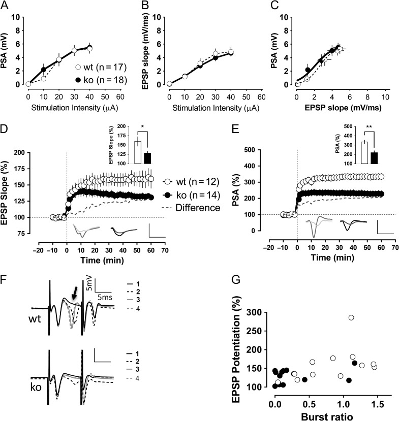
Impaired dentate gyrus synaptic plasticity in Cav3.2 knockout mice. Recordings of evoked field potentials were made after stimulation of the medial perforant path. General excitability and synaptic transmission seemed to be unaffected in knockout animals, as shown in the similar population spike amplitudes (A), EPSP slopes (B) and the EPSP slope relation to the generation of action potentials (C). However, the level of synaptic potentiation recorded 1 h after the TBS was reduced in the knockout group compared with control animals (P < 0.05, Mann–Whitney U-test). In (D and E), the potentiation levels of the field-EPSP and population spike after the TBS protocol are shown. The insets show the potentiation level in the last 10 min of recordings as well as traces corresponding to baseline and 1 h after the TBS. Scale bars: 5 mV/5 ms. (F) During the induction protocol, granule cells tended to fire “bursts” of action potentials, especially after the second repetition of the theta-burst stimulation protocol, as illustrated in the representative traces (upper traces: wild-type slice), whereas the Cav3.2 knockouts were much more reluctant to it (lower traces: Cav3.2 knockout). We quantified the ratio of the second population spike amplitude in the burst to the amplitude of the first spike. The burst ratio in wild-type slices (1.07 ± 0.18) significantly differed from the burst ratio in the knockout group (0.32 ± 0.15) (P < 0.05, Mann–Whitney U-test). (G) This burst ratio was significantly correlated with the actual amount of synaptic potentiation 1 h later (Spearman coefficient of correlation r = 0.55, P < 0.01). *P < 0.05, Mann–Whitney U-test.

