Figure 1.
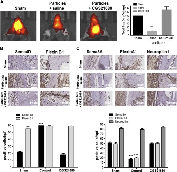
Activation of A2AR increases new bone formation at sites of wear particle-induced osteolysis by changes in axon guidance molecule expression. Mice were administered UHMWPE particles and either treated with saline or the A2AR agonist CGS21680. Control mice (sham) were not exposed to particles (n = 5). Mice were injected daily, and after 2 wk, animals were euthanized. A) XenoLight RediJect Bone Probe 680 conjugate was injected intravenously, and the fluorescence image was captured 1 wk after surgery. Total flux in photons per second was normalized and expressed as a percentage of control to avoid intrinsic changes among animals. Red indicates low-signal intensity and low rates of new bone formation, whereas yellow indicates high-signal intensity and high rates of new bone formation. B) Representative images for Sema4D and PlexinB1 immunohistochemistry and quantification of the number of positive cells/hpf. Cells were counted in 5 different images for each of 5 mice. Data are means ± sem. C) Representative images for Sema3A, PlexinA1, and Neuropilin-1 immunohistochemistry and quantification of the number of positive cells/hpf. Cells were counted in 5 different images for each of 5 mice. Data are expressed as means ± sem (n = 5/group). ***P < 0.001 compared with control (ANOVA). Original magnification, ×40. Original scale bars, 50 µm.

