Figure 2.
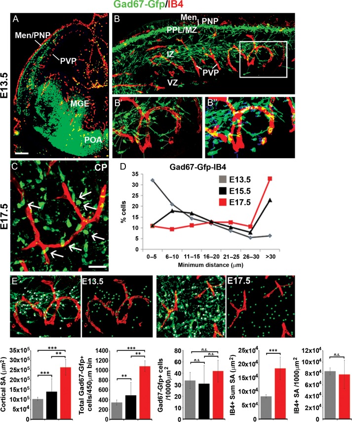
Interneurons are located in close proximity to blood vessels during their tangential and radial phases of migration. (A) Labeling of IB4+ blood vessels, together with immunostaining for Gad67-gfp+ interneurons in the E13.5 forebrain; cortex shown at high magnification in panel (B). (B’–B”) Panels show the acquired confocal image (B’ left panel) with the corresponding false-colored image (B” right panel) showing cells recognized using the bespoke plugin by thresholding according to immunostaining signal intensity and predicted cell size and used to quantify minimum distances between the centroid of migrating interneurons and the closest IB4+ vascular surface. (C) Labeling of IB4+ blood vessels together with immunostaining for Gad67-gfp+ interneurons in the E17.5 mouse cortex, showing radially oriented interneurons close to blood vessels in the CP. (D) Graphs showing the distribution of minimum intracellular distances between Gad67-gfp+ interneurons with IB4+ blood vessels measured using the bespoke plugin in the E13.5 (gray), E15.5 (black) and E17.5 (red) cortex. (E) Panels show confocal images of the E13.5 and E17.5 cortex of Gad67-Gfp+ mouse forebrains with Gad67-gfp+ cells detected using the Imaris software spot module (green spots) and surface rendering of blood vessels (red surfaces) used to quantify interneuron and vascular density. Graphs from left-right show: changes in cortical surface area (μm2), total Gad67-gfp+ interneuron numbers counted in 450 μm cortical bins, interneuron numbers normalized for cortical surface area, IB4+ blood vessel sum surface area, and density of IB4+ blood vessels normalized for cortical surface area at different stages of development (t-test, n = 3 for each; **P ≤ 0.01, ***P ≤ 0.001). (Men/PNP, Meninges/perineural plexus; PVP, periventricular plexus; MGE, medial ganglionic eminence; POA, preoptic area; MZ, marginal zone; CP, cortical plate; SP, subplate; IZ, intermediate zone; LIZ/SVZ, lower intermediate zone/subventricular zone; VZ, ventricular zone).

