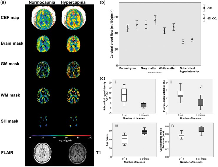
Figure 2.
Cerebral blood flow and vasoreactivity – ASL MRI. (a) Generated CBF maps were masked with brain, grey matter, white matter and subcortical hyperintensity masks and the average in each mask recorded. Masks were created using T1 and FLAIR images. Scale bar shown for CBF in ml/100 g/min (b) CBF in brain, grey matter, white matter and subcortical hyperintensities whilst breathing air (line) and 6% CO2 (dashed line). (c) Patients with five or more lacunes had (i) lower cerebral vasoreactivity and (ii) lower peripheral vasoreactivity (brachial FMD). There were also (iii) older and had higher carotid intima-media thickness (iv). Boxplot shows medians, quartiles, and extreme values.
CBF: cerebral blood flow; GM: grey matter; WM: white matter; SH: subcortical hyperintensity.

