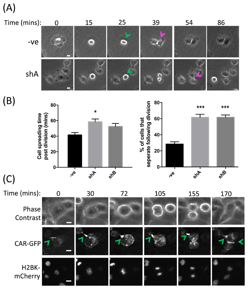
Figure 2. CAR promotes post-mitotic daughter cell attachment and spreading.
(A) Representative images from time-lapse movies of shControl or CAR-KD (shA) A549 cells undergoing division. Green arrows denote dividing cells, red arrows denote initiation of daughter cell separation following division. (B) Quantification of time-lapse movies of control and CAR-KD cells (shA, shB) represented in (A), assessing the time taken for cells to re-spread post-division (left) and the percentage of cells that separate completely after division (right). Data are quantified from at least 20 cells over 3 independent experiments. Data are mean ± SEM. *p<0.05, ***p<0.005. (C) Representative images from time-lapse movies of A549 cells expressing CAR-GFP and H2BK-mCherry; n = 4 experiments. Green arrows denote sites of high CAR-GFP at cell-cell contact points. Scale bars are 10um.

