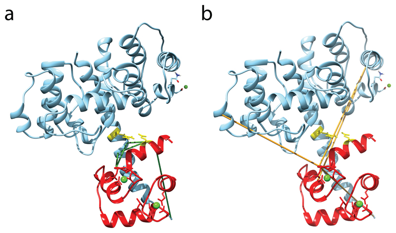
Figure 5. Predicted structure of the plectin ABD–calmodulin complex.
(a,b) Compatible (a) and incompatible (b) cross-links mapped onto the selected model of the complex between plectin ABD and the N-terminal lobe of calmodulin. The calmodulin chain is colored red. Compatible and incompatible cross-links are colored green and orange, respectively, and calcium and magnesium ions are shown as green balls. Residues E14 of calmodulin and R40 of plectin, the residues involved in the known salt bridge formation, are shown as sticks and are colored yellow.

