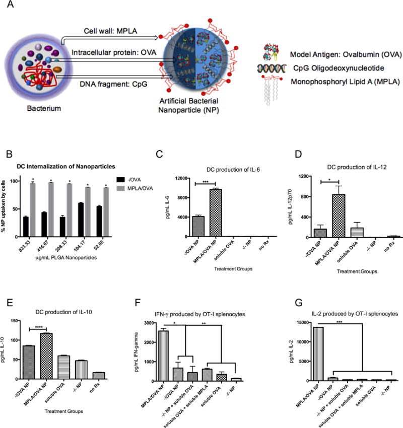
Figure 1.

MPLA on the surface of NP, mimicking bacterial cell walls (A), increases BMDC uptake, pro-inflammatory cytokine production, and antigen-specific T cell proliferation. (B) BMDCs incubated with titrated PLGA NP internalize MPLA-coated particles more avidly than blank NP (*p<.0001 for all concentrations) (C) After 24h with NP, BMDCs produce a higher amount of pro-inflammatory cytokines IL-6 (***p=.0001), (D) IL-12 (*p=.0217), and (E) IL-10 (****p<.0001) in response to MPLA/OVA vs. −/OVA NP. (F) Co-incubating OVA-specific OT-I splenocytes with NP-treated BMDCs results in greater production of proliferative cytokines IFN-gamma (*p<.05, **p<.01) (G) and IL-2 (****p<.0001) after 5 days. Results are representative of three independent experiments. Error bars represent Standard Experimental Error (SEM).
