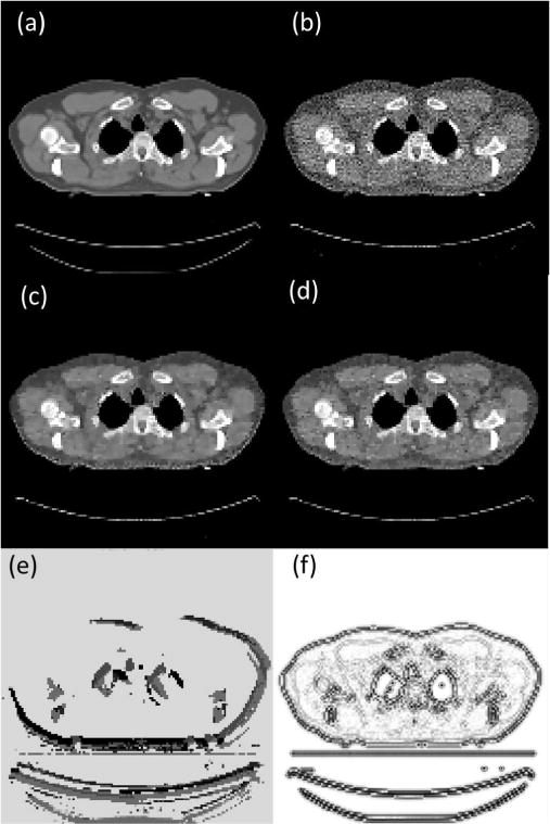
Fig. 7.

(a) Ground truth CT image of a case that is used in training PTPN. (b) Image reconstructed with an arbitrarily selected parameter λ(x) = 0.005. (c) Image reconstructed after the parameter is tuned by PTPN. (d) Image reconstructed with manually tuned λ(x) = 0.05. CT images are displayed in the window [−100, 300] HU. (e) Tuned parameter map λ(x) displayed in log10 scale. (f) Optimal parameter map λ∗(x).
