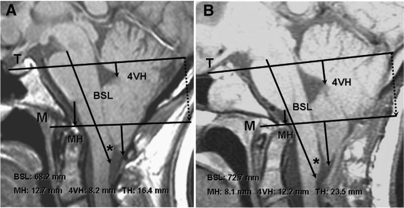
Fig. 2.
Morphometric assessments of CCJ in 24-year-old woman with CM-I/TCS before and after failed Chiari surgery. A: Preoperative midsagittal T1-weighted MR image showing elongation of brain stem (BSL = 68.2 mm), downward displacement of medulla (MH = 12.9 mm), downward displacement of cerebellum (4VH = 7.6 mm), and herniation of cerebellar tonsils (TH = 17.8 mm). To reconstruct line M on postoperative films, the distance between the internal occipital protuberance and line M was measured along a line drawn perpendicular to line T (dotted line). B: postoperative scan with reconstructed line M, 6 months after posterior fossa decompression showing cerebellar ptosis with greater elongation of brain stem (BSL = 72.7 mm), greater downward displacement of medulla (MH = 7.6 mm), greater downward displacement of cerebellum (4VH = 12.9 mm), and greater herniation of cerebellar tonsils (TH = 25.0 mm). Asterisk indicates gracile tubercle.

