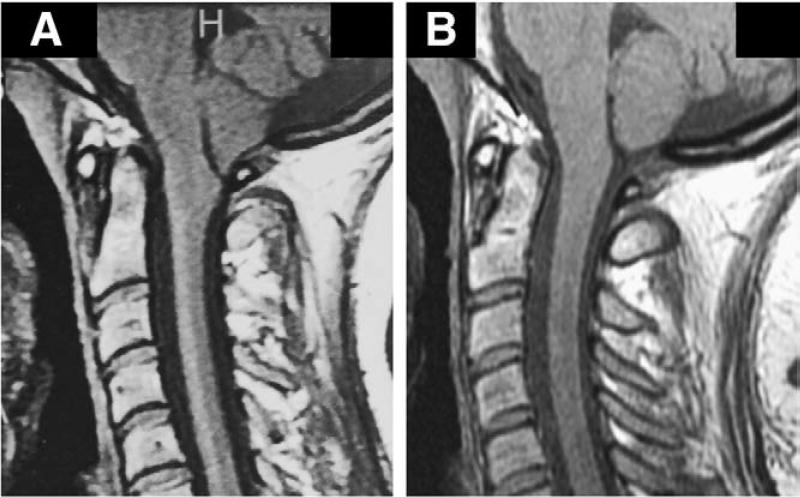
Fig. 6.
Midsagittal T1-weighted MR images before and after SFT in 28-year-old woman with CM-I/TCS. A: Preoperative scan showing tonsillar herniation extending to superior arch of C1. Morphometric measurements revealed elongation of brain stem (BSL = 60.1 mm), downward displacement of medulla (MH 7.7 mm), downward displacement of cerebellum (4VH = 14.4 mm), herniation of cerebellar tonsils (TH = 12.3 mm), and enlargement of FM (transverse diameter = 34.3 mm). B: Postoperative scan 7 months after SFT showing resolution of CM-I. Morphometric measurements revealed normalization of brain stem length (BSL = 53.7 mm), resolution of hindbrain displacement (MH = 12.4, 4 VH = 4.0 mm), and ascent of cerebellar tonsils (TH = 1.5 mm).

