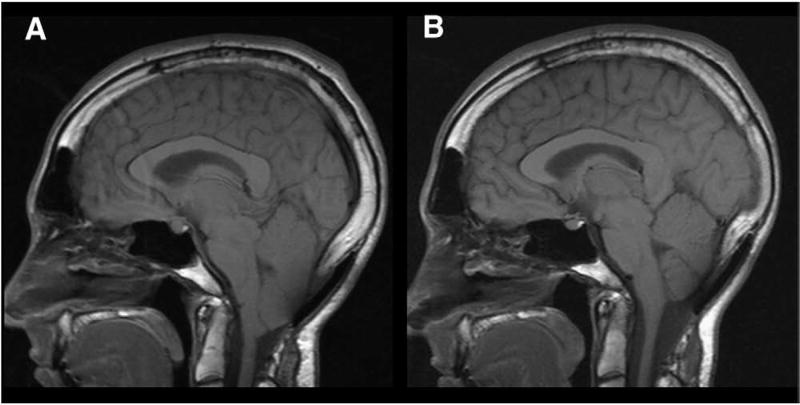
Fig. 8.
Midsagittal T1-weighted MR images before and after SFT in 27-year-old man with CM-I/TCS who developed progressive cerebellar ptosis and recurrent Chiari symptoms after posterior fossa decompression. A subsequent cranioplasty 1 year before SFT failed to arrest ongoing prolapse and clinical deterioration. A: Preoperative scan showing suboccipital craniectomy, overlying cranioplasty plate, and cerebellar ptosis with elongation and downward displacement of hindbrain. The cerebellum is grossly misshapened and vertically oriented. Morphometric measurements confirmed elongation of brain stem (BSL = 61.7 mm), downward displacement of medulla (MH = 10.8 mm), and downward displacement of cerebellum (4VH = 28.7 mm). B: Postoperative scan 8 months after SFT showing resolution of cerebellar ptosis and anatomical remodeling of cerebellum. Morphometric measurements revealed normalization of brain stem length (BSL = 52.8 mm) with ascent of medulla (MH = 14.4 mm) and cerebellum (4 VH = 18.1 mm).

