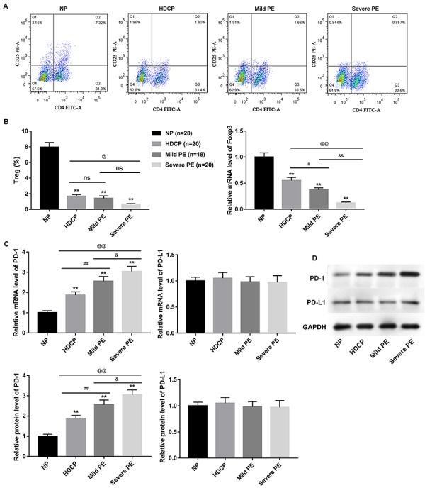
Figure 2. Expression of Treg and Foxp3 as well as PD-1/PD-L1 expression in Treg cells. Peripheral blood mononuclear cells were isolated from the venous blood of women with normal pregnancy (NP), hypertensive disorder complicating pregnancy (HDCP), mild preeclampsia (PE), and severe PE by density gradient centrifugation. A, The percentage of Treg cells was assessed by flow cytometry. B, The mRNA level of Treg-specific transcript factor Foxp3 was evaluated by qRT-PCR. C and D, The mRNA and protein levels of PD-1 and PD-L1 in CD4+CD25+ Treg cells were assessed by qRT-PCR and western blot, respectively. Data are reported as means±SD. **P<0.01 vs NP; #P<0.05, # #P<0.01 mild PE vs HDCP; @P<0.05, @@P<0.01 severe PE vs HDCP; &P<0.05, &&P<0.01 mild PE vs severe PE; ns: not significant (t-test).

