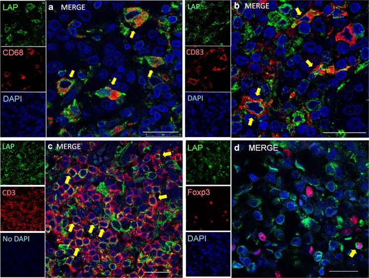
Fig. 3.
Double immunofluorescence for LAP (TGFβ1) (green) plus CD68 (red)(a), CD83 (red)(b), CD3 (red)(c), and FoxP3 (red)(d) by confocal laser scanning microscopy. Arrows, double positive cells. LAP (TGFb1)+ immune cells include macrophages (a) (most abundant), mature conventional dendritic cells (cDCs) (b), and a part of T cells (c). It is noteworthy that Treg cells (d) infrequently co-express LAP (TGFβ1). Scale bars, 10 μm (a–d)

