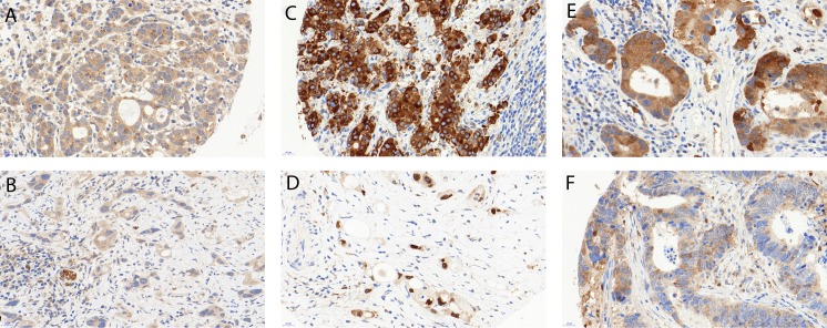
Fig 1. Examples of immunohistochemical stainings.
(a) High LC3B dot like staining (score 2). (b) Low LC3B dot like staining (score 1), note a small nerve serving as internal positive control. (c) High p62 cytoplasmic staining (score 3), while negative nuclear staining. (d) Low p62 cytoplasmic/dot-like staining (scores 0), positive nuclear staining. (e) High cytoplasmic (score 2) and low dot-like (score 1) p62 staining. (f) Low cytoplasmic (score 1) and high dot like (score 2) p62 staining. 40x magnification for all images. Error bars indicate 20µm.

