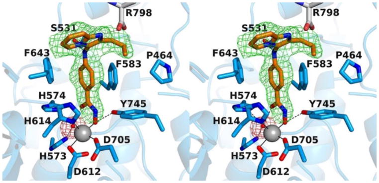
Figure 2.
Polder omit maps (stereoview, contoured at 3.0 σ each) for 4l (green) and the Zn2+-bound water molecule (magenta) in the active site of HDAC6. Atom color codes are as follows: C = orange (4l) or light blue (protein), N = blue, O = red; Zn2+ appears as a grey sphere, and the Zn2+-bound water molecule is shown as a small red sphere. Metal coordination and hydrogen bond interactions are indicated by solid and dashed black lines, respectively.

