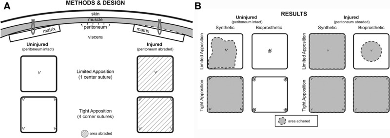
Fig. 2.

A, Study design for implantation of mesh into rat peritoneum. Through a vertical midline incision, 2 pieces of mesh were placed within each animal. Care was taken in avoid any suture remaining intraperitoneally to avoid adhesion and/or vascular contributions from the viscera. The peritoneum was sharply abraded where indicated. Animals were evaluated after 5 weeks. B, Schematic of Results. The synthetic mesh was typically adherent under any condition. The bioprosthetic mesh was adherent only at the areas of suture fixation when peritoneal lining was left intact, whereas adherence was significantly greater in the areas of denuded lining.
