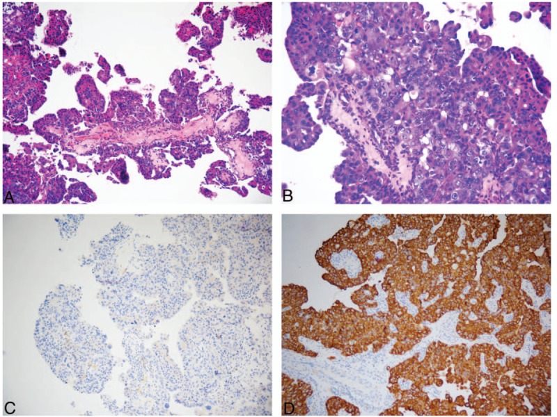
Figure 4.

Pathological characteristics and immunohistochemical findings in the tumor in the third surgery. (A) Low-power view showing the predominantly papillary surface pattern of the tumor and cell congestion. (B) Medium-power view showing an increase in number of cell layers, and the cells were crowded and heterotypic. (C) Negative staining for the CK5/6 squamous cell carcinoma markers. (D) Positive staining for the CK7 adenocarcinoma markers.
